3D Cell Culture Models: Techniques, Applications, and Best Practices
Creating 3D cell culture models has transformed how I approach biological research and visualization. These models provide a more realistic environment for studying cellular behavior, which is crucial for applications like drug testing and disease modeling. In this article, I’ll break down the practical steps, tools, and best practices I use to generate accurate and production-ready 3D cell culture models. Whether you’re a researcher, designer, or developer, you’ll find actionable advice for optimizing your workflow and avoiding common pitfalls.
Key takeaways:
- 3D cell culture models offer more realistic biological insights than 2D cultures.
- AI-powered tools streamline segmentation, retopology, and texturing.
- Careful workflow planning avoids errors and speeds up production.
- Applications range from drug discovery to regenerative medicine.
- Collaboration and scaling require clear protocols and tool compatibility.
Understanding 3D Cell Culture Models
What are 3D cell culture models?
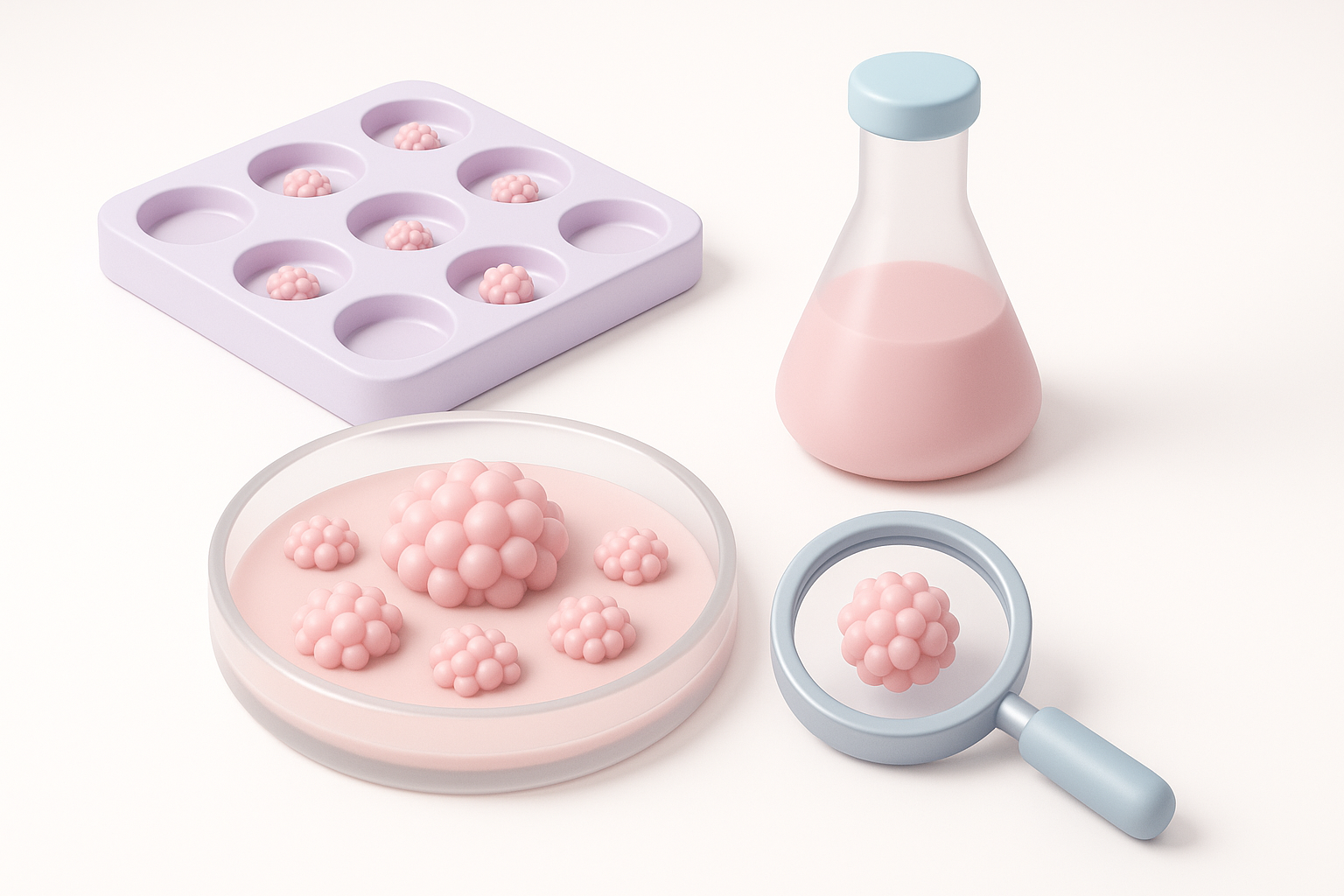
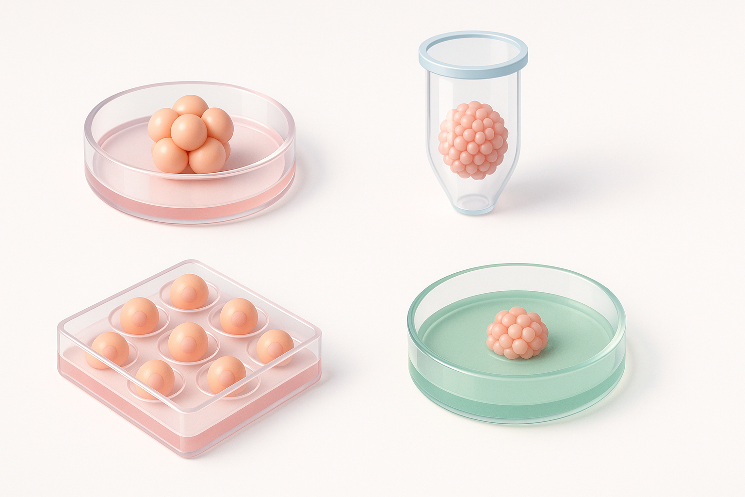
3D cell culture models are in vitro systems where cells grow in a three-dimensional environment, more closely mimicking the architecture and interactions found in living tissues. Unlike flat, two-dimensional cultures, these models enable cells to interact spatially, which affects their morphology, gene expression, and behavior.
I often use scaffold-based, spheroid, or organoid approaches, depending on the biological question. These methods let me observe complex cell–cell and cell–matrix interactions, which are critical for realistic modeling.
Key differences from 2D cell cultures
The main difference is dimensionality: 2D cultures restrict cells to a flat surface, while 3D models allow cells to grow and interact in all directions. This leads to:
- More physiologically relevant gene and protein expression.
- Better simulation of tissue-like gradients (e.g., oxygen, nutrients).
- Improved prediction of in vivo drug responses.
In my experience, moving to 3D models reveals cell behaviors that are completely missed in 2D, leading to more robust and translatable results.
My Workflow for Creating 3D Cell Culture Models
Step-by-step process
Here’s my typical workflow for generating a 3D cell culture model:
- Data Collection: Gather reference images or sketches from microscopy or literature.
- Initial 3D Generation: Use an AI-powered platform like Tripo to convert these references into a base 3D mesh.
- Segmentation: Automatically or manually segment cellular components (nuclei, cytoplasm, ECM).
- Retopology: Clean up the mesh for optimal geometry and downstream processing.
- Texturing: Apply realistic textures to highlight cellular features.
- Validation: Compare the model to biological data for accuracy.
This streamlined process minimizes manual labor and reduces errors.
Tools and platforms I use
I rely on AI-driven platforms for most of the heavy lifting. Tripo, for example, lets me input images or sketches and outputs a segmented, retopologized 3D model ready for texturing. For fine-tuning, I use standard 3D editing software and visualization tools that support scientific formats.
Checklist:
- Ensure input data is high-quality and representative.
- Use automated segmentation where possible to save time.
- Always validate against experimental data.
Best Practices for 3D Cell Culture Model Generation
Optimizing segmentation and retopology
Accurate segmentation is key for biologically meaningful models. I recommend:
- Using AI-assisted segmentation to distinguish cellular structures.
- Manually correcting any errors in critical regions.
- Running retopology tools to ensure the mesh is clean and efficient for rendering or simulation.
Pitfalls to avoid:
- Over-segmentation, which can create unrealistic boundaries.
- Skipping retopology, leading to heavy, unmanageable files.
Ensuring accurate texturing and visualization
Texturing brings biological realism to your models. I usually:
- Reference real microscopy images for texture maps.
- Use procedural texturing to simulate subcellular variability.
- Test visualizations in different lighting conditions to ensure clarity.
Tips:
- Keep textures biologically plausible.
- Check that textures scale correctly with model resolution.
Applications and Use Cases in Research and Industry
Drug discovery and testing
3D cell culture models are now standard in preclinical drug screening. They better mimic tissue responses, leading to more predictive results. I’ve seen improved accuracy in cytotoxicity and efficacy studies when using 3D models versus traditional 2D assays.
Best practices:
- Validate drug responses with both 2D and 3D systems for comparison.
- Use automated platforms to handle large sample sets efficiently.
Disease modeling and regenerative medicine
For disease modeling, 3D cultures allow me to recreate disease microenvironments (e.g., tumor spheroids, fibrotic tissue). In regenerative medicine, these models support stem cell differentiation and tissue engineering by providing the right spatial cues.
Use case tips:
- Integrate patient-derived cells for personalized models.
- Regularly update models as new biological data emerges.
Comparing 3D Cell Culture Modeling Methods
AI-powered vs traditional workflows
AI-powered workflows, like those with Tripo, dramatically reduce manual steps:
- Faster segmentation and retopology.
- Consistent output quality across projects.
- Lower barrier for non-specialists.
Traditional workflows require more manual modeling and expertise, which can slow down projects and introduce variability.
Decision guide:
- Use AI-powered tools for rapid prototyping and standard tasks.
- Reserve manual workflows for highly specialized or novel structures.
Choosing the right approach for your project
I assess:
- Project complexity (simple vs. complex tissue structures).
- Team expertise (AI tools are more accessible for newcomers).
- Scale (AI workflows scale better for large studies).
Checklist:
- Match tool capabilities to project needs.
- Factor in validation and review time for critical applications.
Tips and Lessons Learned from Real-World Projects
Common challenges and how I solve them
Challenge: Inaccurate segmentation of overlapping cells
Solution: Combine AI-based segmentation with manual correction for key regions.
Challenge: Large file sizes slow down rendering
Solution: Use retopology and mesh simplification tools before exporting.
Challenge: Inconsistent textures
Solution: Standardize texture sources and test across multiple visualization platforms.
What I’ve learned about scaling and collaboration
Scaling up requires:
- Consistent file naming and version control.
- Clear documentation of model generation steps.
- Using platforms that support team collaboration and cloud storage.
Collaboration tips:
- Share intermediate models for feedback early.
- Use platforms compatible with common scientific and 3D formats.
By following these best practices and leveraging AI-powered tools, I’ve significantly improved both the speed and quality of my 3D cell culture modeling projects. Whether you’re just starting or looking to optimize your workflow, these strategies will help you achieve reliable, reproducible results.


